
Contact us right now
X-ray
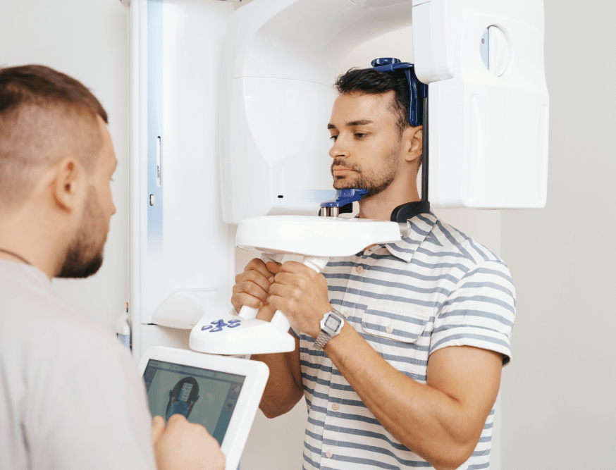
SIA “Klīnika Denta” has equipped its X-ray room with a new state-of-the-art Carestream 3D computed tomography scanner to provide our patients with the highest level of dental diagnostics.
With this device we are able to perform the following X-ray diagnostics:
- 3D jaw computed tomography for both jaws, each individually or by segments.
- Digital orthopantomogram (panoramic X-ray).
- Supplementary sinus X-ray.
X-ray
SIA “Klīnika Denta” has equipped its X-ray room with a new state-of-the-art Carestream 3D computed tomography scanner to provide our patients with the highest level of dental diagnostics.
With this device we are able to perform the following X-ray diagnostics:
- 3D jaw computed tomography for both jaws, each individually or by segments.
- Digital orthopantomogram (panoramic X-ray).
- Supplementary sinus X-ray.
ADVANTAGES
Thanks to 3D images, our dentists and implantologists are able to accurately plan the implant placement site before inserting a dental implant, because unlike a conventional X-ray, a 3D X-ray permits visualizing and measuring bone thickness, depth and distance to the mental nerve and sinuses. For more complex surgeries,
implant dentists are able to digitally simulate implant placement, thus reducing the risk of errors during surgery.
Advantages
Thanks to 3D images, our dentists and implantologists are able to accurately plan the implant placement site before inserting a dental implant, because unlike a conventional X-ray, a 3D X-ray permits visualizing and measuring bone thickness, depth and distance to the mental nerve and sinuses. For more complex surgeries, implant dentists are able to digitally simulate implant placement, thus reducing the risk of errors during surgery.
Why dental X-rays are necessary and how are they different?
Patients often come to the dentist with dental X- rays that have been taken a year ago or even earlier. To make an accurate dental diagnosis, it is essential to see what is the current situation in the mouth. In one year, a tooth can easily change from healthy to damaged. Tooth decay can start between the teeth or in the root of the teeth where the canals were Ylled, or cysts may also form. Therefore, the X-rays should be no older than 6 months.

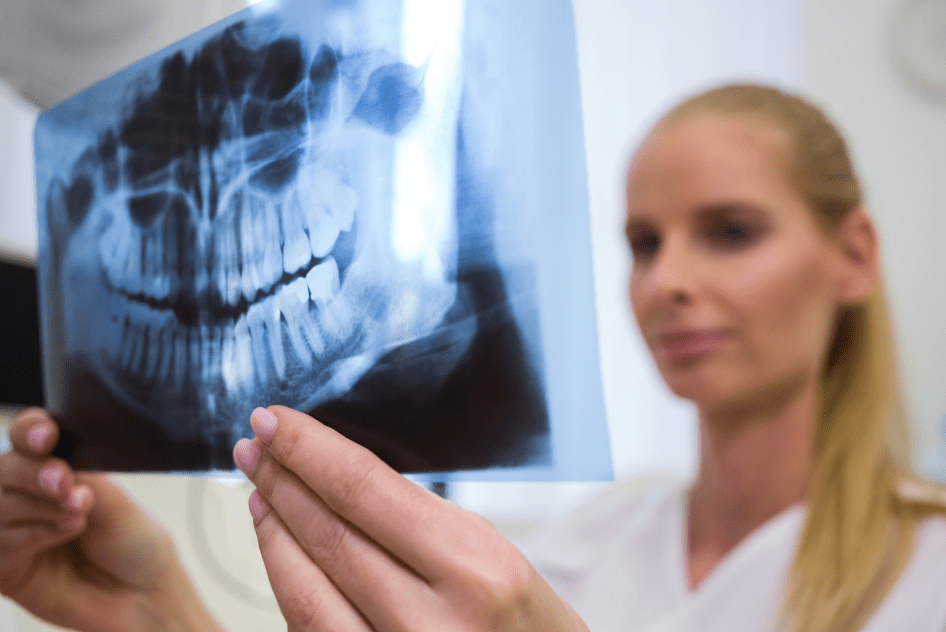
2D X-ray
Another issue with conventional (Ylm or digital sensor) X-rays is that they provide little information and can help diagnose the condition of 1-2 teeth. Therefore, it is di[cult to draw up an accurate treatment plan. A panoramic dental X-ray is a two-dimensional, 2D X-ray image, hence, there are distorted areas, some are stretched and others are overlapping.
Consequently, it is not always possible to see additional root canals or in\ammation at the apex of the root. It is also not possible to assess bone thickness and density before implant placement. Panoramic X-rays allows general information about the oral cavity to be obtained.
3D X-ray
A 3D X-ray is a three-dimensional X-ray image, the 3D system X-ray is displayed in three-layer projections, without any distortion, in a 1: 1 ratio. Accordingly, it is possible to assess the condition of teeth and tissues with surgical precision. Therefore, the patient needs a 3D X-ray for their own beneYt.